

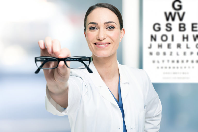


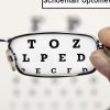

About Pasteur Eye Hospital
Pasteur Eye Hospital (previously Horizon Eye Care Centre) is a modern, dedicated and fully equipped eye hospital situated in the rustic suburb of Hospital Park in Bloemfontein. Located at 54 Pasteur Drive, we offer a comprehensive range of quality ophthalmic services. We provide holistic eye care treatments with therapeutic and diagnostic research and development components to benefit our patients.
We provide ophthalmic examinations and eye care to patients in the Free State, Northern and Eastern Cape. Our team of highly specialized and experienced ophthalmologists is supported by a team of dedicated ophthalmic nurses.
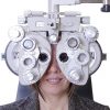
Pasteur Eye Hospital provides a complete range of eye care services, including visual field testing, optical coherence tomography, ophthalmoscopy, fluorescein angiograms, and PRK and Lasic surgery.
Your eyes are the windows to your health
For centuries, the literary world and artists alike have used the metaphor “windows to the soul” to indicate how important the eye is to the rest of the body. It is described as a first point of contact, as it were.
When a patient arrives at Pasteur Eye Hospital with a certain problem, the eye is a treasure trove of wealth to be examined. It holds opportunities and clues for the practitioners in the medical science to detect and successfully diagnose illnesses.
What an eye examination reveals
The retina is the inner, thin layer of tissue that lines the back of the eye. Using special technology, such as a split lamp with specialized lenses, a retinal camera, or even doing an OCT (Optical Coherence Tomography), an Ophthalmologist (eye specialist) can visualize the retina with clarity. When a change on the retina is observed, further tests can be done in cases where Diabetes, Hypertension, and High Cholesterol are suspected. There are certain marks that are left of the retina by parasitic infections such as Toxoplasmosis, Toxocara and even Malaria.
The eye is so revealing, even the use of certain medications can leave their mark on the retina. Some of these include steroids, anti-malaria medicines, and medicines that treat heart conditions.
The close relation between the eye and the brain enables the development of new and innovative technology which can assist with a number of conditions. Increased pressure in the brain can be detected by scanning the back of the eye.
The eye assists in the detection of serious conditions, such as cranial deformities, trauma suffered, infections as well as hydrocephalus. The latest technology in diagnostics through the eye, is an ultrasonic machine which tracks the dynamics of the back of the eye. When monitoring these dynamics, the amount of pressure in the brain can be determined.
At Pasteur Eye Hospital, we take pride in our dedicated complement of specially trained staff, our highly qualified and experienced ophthalmologists and our cutting-edge equipment.
Book an appointment with one of the specialist practitioners at Pasteur Eye Hospital today.
Please do not hesitate to contact us for more information or to make an appointment.
Eye Care Services at Pasteur Eye Hospital
In addition to standard eye examinations and ophthalmic care, our skilled ophthalmologists provide a variety of special examinations in our dedicated examination area. This means that you don’t have to be referred to a different eye specialist or facility. Everything is taken care of by our team, making Pasteur Eye Hospital is your one-stop centre for eye care excellence.
Our special eye examinations include:
Fluorescein angiograms
Used to examine pathology of the retina. The patient’s eye is injected with a dye called Fluorescein. This colours the blood vessels in the eye. By means of photography, any leakage is picked up. The leakage will then be treated.
Corneal topography
This test is used to provide information on the cornea, specifically on its curvature. We have a computer program which analyse the cornea surface. A colour interpretation map helps the ophthalmologist pick up corneal diseases and irregularities and to correct astigmatism. It is also an important examination to provide measurements for establishing data to calculate the power of intraocular lenses and refractive surgery.
Fundus photography
Fundus photography is used to establish the condition of the retina, macula and optic nerve. Diseases like glaucoma and macular degeneration can be tracked by this method
Keratometry
This is used to describe the measurement of the hills and valleys of the cornea. Again this information is used to determine the power of the intraocular lens to be used.
Ophthalmoscopy
This method is used by the doctor to examine the retina and vitreous of the eye. He uses an ophthalmoscope (direct or indirect) to perform this test.
Gonioscopy
This is usually part of the examination for glaucoma. It examines the anterior chamber angle – that is the part of the eye that directs the aqueous humor produced by the eye.
Optical Coherence Tomography
The abbreviation for this examination is the so called OCT. By using this technique one can get cross-sectional images of the retinal tissue. BY means of this technique, early signs of defects of the retina can be detected and therefore treated timeously. Important diseases picked up with this technique are macular degeneration, retinal diseases and glaucoma. Also useful for providing information for refractive surgery (Excimer laser).
Tonometry
This technique is used to measure the pressure within the eye. Useful in screening patients for glaucoma.